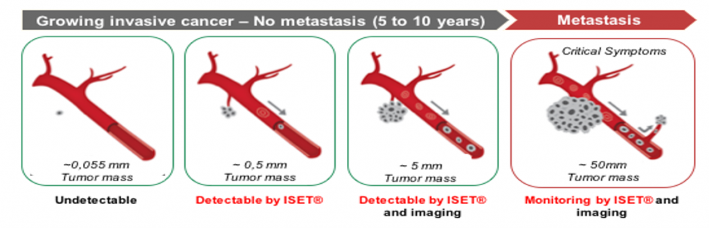
The earlier CTC are detected, the faster and more effectively a patient can be treated. This non-invasive approach could lead to efficient screening test for population at risk such as heavy smokers.
Ilie et al. 2014 Plos One
Centre Hospitalier Universitaire De Nice
An independently funded, 6-year-long monocentric study, published in 2014, demonstrated that ISET® was able to make a very early diagnosis of lung cancer.
254 patients including 168 patients with COPD followed by ISET® and low dose CT-scan screening :
5 of 168 presented CTCs detected by ISET®.
Per 10 mL of blood, 19 to 67 isolated CTCs and 1 to 3 CCMs, Vimentin expression.
In these 5 patients :
CT-scan detected a lung nodule 1 to 4 years after CTCs detection by ISET® leading to surgical resection and early diagnosis of Lung Cancer (stage 1A).
Follow up of the 5 patients at 18 months after surgery: no tumor recurrence, no CTC.
National Institute of Integrative Medicine
542 individuals tested with ISET®: 277 patients with cancer and 256 individuals at risk of developing cancer :
100% of cancer patients presented CTC.
132 at risk individuals (50%) presented CTC.
10 months follow-up of CTC-positive at risk individuals :
20% of at risk individuals diagnosed with early stage cancers by imaging techniques.
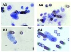
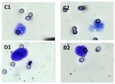
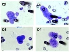
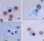
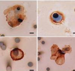
Images below present cytomorphological detection of CTCs using the ISET method. (A) Breast cancer, (B) Prostate cancer, (C) Colorectal cancer, (D) Renal/bladder cancer.
Ried et al. 2017 Asian Pacific Journal of Cancer Prevention
Ried et al. 2017 Asian Pacific Journal of Cancer Prevention
542 individuals tested with ISET®: 277 patients with cancer and 256 individuals at risk of developing cancer :
100% of cancer patients presented CTC.
132 at risk individuals (50%) presented CTC.
10 months follow-up of CTC-positive at risk individuals :
20% of at risk individuals diagnosed with early stage cancers by imaging techniques.
Images below present cytomorphological detection of CTCs using the ISET method. (A) Breast cancer, (B) Prostate cancer, (C) Colorectal cancer, (D) Renal/bladder cancer.
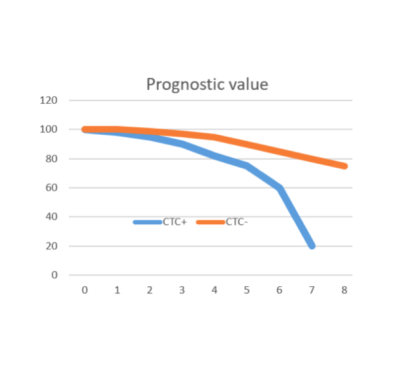
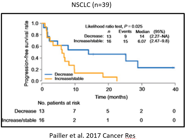
The prognostic value of CTC collected by ISET® has been demonstrated by multiple independent publications in the context of multiple cancer types
Prognosis value of CTC in multiple cancers
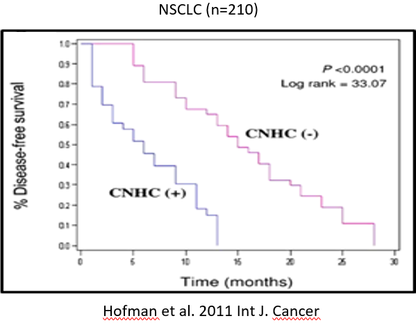
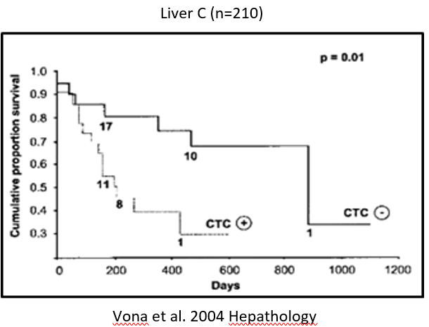
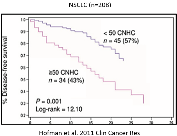
The prognostics value of CTCs enriched by the ISET method has been demonstrated in the context of multiple cancers, such as Hepato-Cellular Carcinoma, Non-Small-Cell Lung Cancer, Small-Cell Lung, Uveal Melanoma, Colorectal Cancer, Pancreatic Cancer, Head and Neck, Ovarian Cancer and Soft Tissue Sarcoma.
Examples of Kaplan-Meyer curves disease-free survival duration stratified according to the presence or absence of CTCs are presented in the images below.
Theranostics allows tailoring to individual-patients new targeted treatments. These treatments are selected based on the molecular characteristics of cancer cells, thus allowing predictably-obtain cancer’s response to treatment. Theranostics markers are commonly used on tissue biopsies (primary tumors, metastasis). The ISET® method is non-invasive and can be performed repeatedly, therefore Circulating Cancer Cells and Microemboli isolated by ISET® allows the follow-up of cancers and the tailoring of the treatment based on real-time analyses of the evolving tumor cells-population.
CTC and ALK status
Positive Control (A549) of a 3-colors -immunofluorescence (Dapi-blue/N-cadherin red/E-cadherin green)
18 ALK-positive patients
14 ALK-negative patients
100% consistent result
100% of patients with detectable CTCs by ISET®
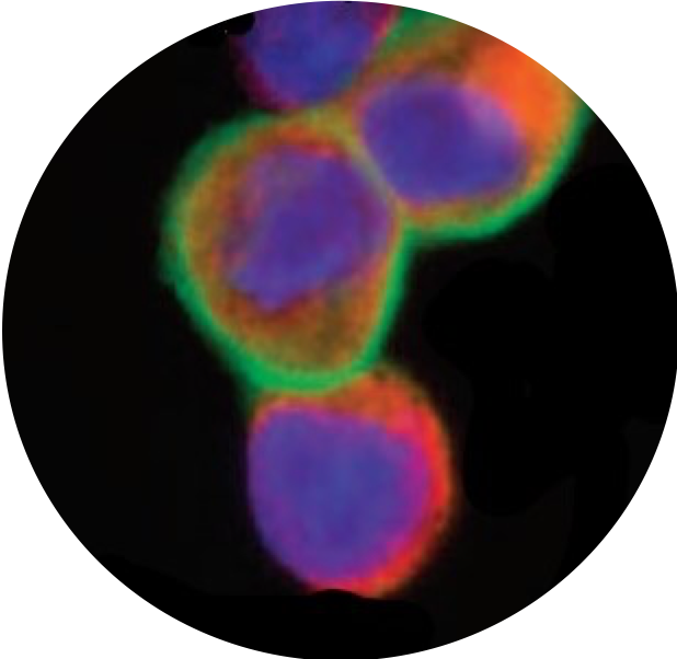
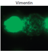
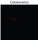
Single cell analysis: ALK-rearranged CTCs expressed a mesenchymal phenotype contrasting with heterogeneous epithelial and mesenchymal marker expressions in tumors. ALK-rearranged CTCs harbored a unique split pattern, and heterogeneous patterns of splits were present in tumors.
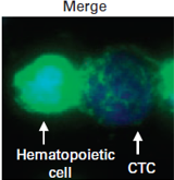
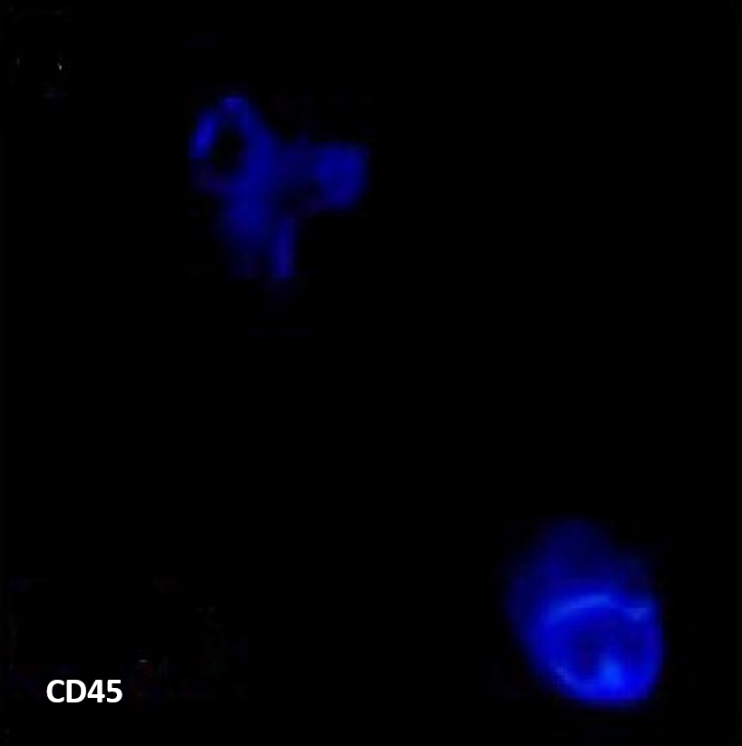
Below is a representative example of vimentin/cytokeratins/CD45/DAPI immunofluorescent staining of ALK-rearranged CTCs in an ALK-positive patient (Paillet et al. 2013 J Clin Oncol).
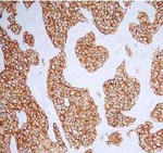
aberrant MET Expression
256 non-small-cell lung cancer patients (stage III-IV)
106 lung adenocarcinoma patients (stage III-IV)
CTC detected by ISET in 75% of patients (all stages)
93% concordance of MET expression on CTC and matched patient tissue
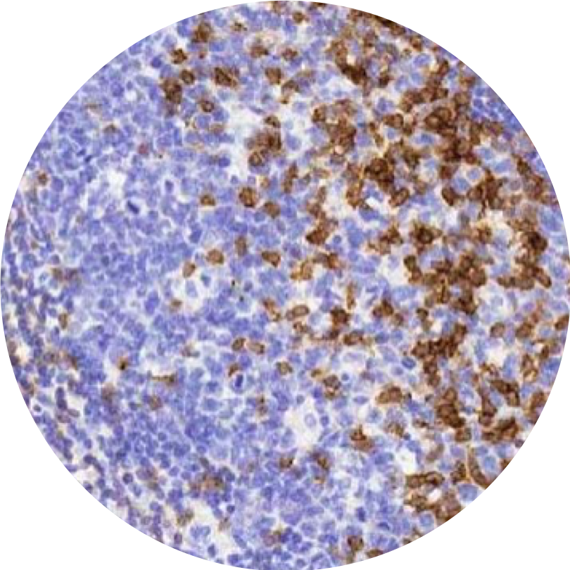
MET protein staining in tumor tissue and corresponding CTCs from selected NSCLC patients, are shown in images examples below.
Ilie et al. 2017 Oncotarget
Ilie et al. 2017 Oncotarget
256 non-small-cell lung cancer patients (stage III-IV)
106 lung adenocarcinoma patients (stage III-IV)
CTC detected by ISET in 75% of patients (all stages)
93% concordance of MET expression on CTC and matched patient tissue
MET protein staining in tumor tissue and corresponding CTCs from selected NSCLC patients, are shown in images examples below
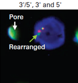
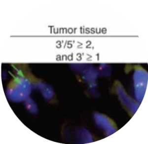
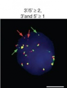
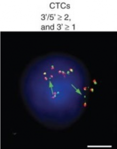
Rearrangement of ROS1
Pailler et al. 2015 Ann Oncol
8 metastatic non-small-cell lung cancer patients
4 ROS1-rearranged tumors
ROS1 rearrangement was detected in CTCs of all 4 patients with ROS1-rearranged primary tumor
Images below show the detection of ROS1-gene alterations in CTCs from RO1-rearranged patients.
The progress of immune surveillance and especially immune evasion in cancer cells are driving promising results for treating cancer with efficacy and low toxicity using immunotherapy. The effect of this treatment could be directly followed and adjusted using the non-invasive ISET technology repeatedly.
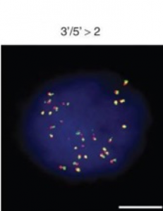
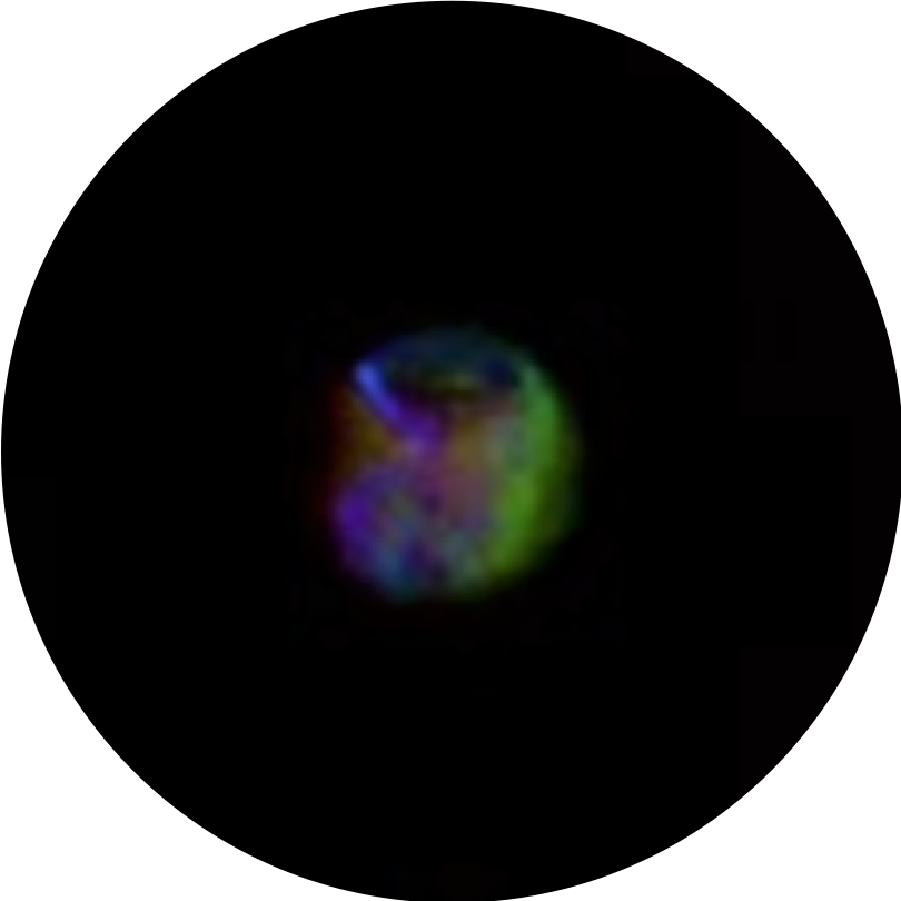
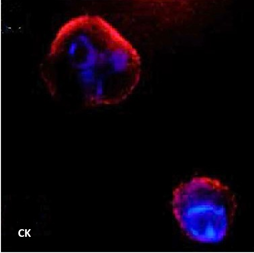
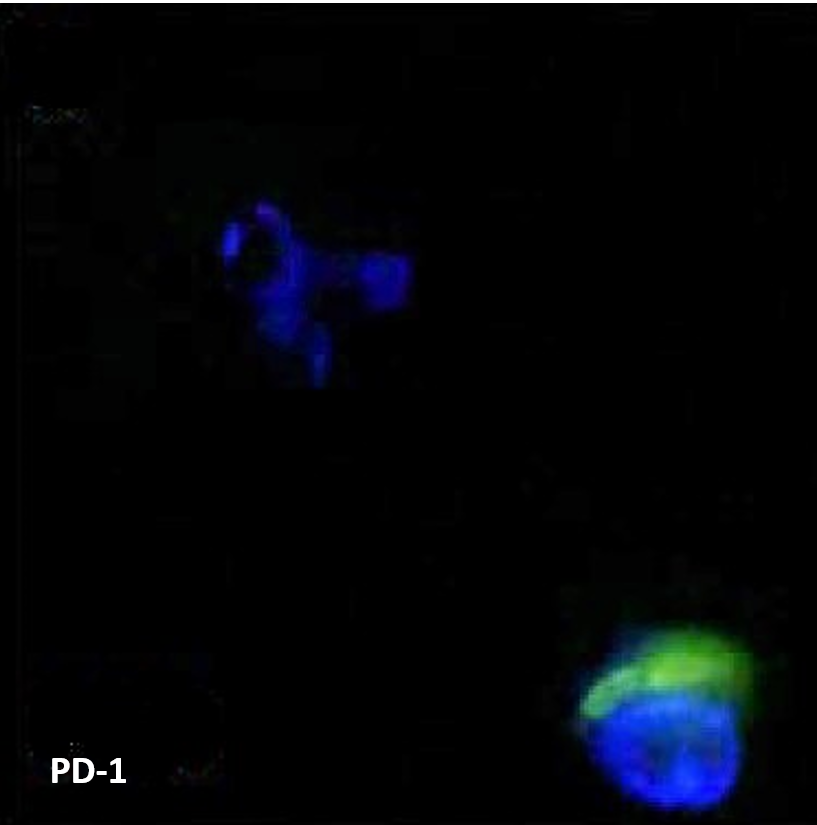
Evaluation of PD-L1/PD-1 on circulating tumor cells
ARIOL image of CTC stained for PD-L1 orange /CK green/CD45 far red
30 metastatic non-small-cell lung cancer patients
Cytopathological analysis identified CCC in 28 patients (93,3%)
By CK+/CD45- IF, epithelial CTC were detected in 17 patients (56,7%)
Median progression-free survival (PFS) was significantly shorter in patients with >3 PD-1(+) CTCs at baseline compared with those with <3 PD-1(+) CTCs (p = 0.022) as well as in patients with >1 Giemsa-positive tumor cells (p = 0.025).
The images below show the detection of PD-L1 in circulating tumor cells and white blood cells (Kallergi et al. 2018 Ther Adv Med Oncol).
Detection of PD-L1 in circulating tumor cells and white blood cells
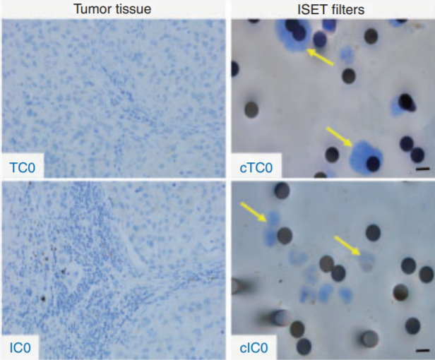
106 lung adenocarcinoma patients (stages III-IV)
Cytopathological analysis identified CCC in 80 patients (75%)
93% concordance of PD-L1 expression between tissue and CTCs (Sensitivity=55%; Specificity=100%)
Ilie et al. 2017 Ann Oncol
Ilie et al. 2017 Ann Oncol
106 lung adenocarcinoma patients (stages III-IV)
Cytopathological analysis identified CCC in 80 patients (75%)
93% concordance of PD-L1 expression between tissue and CTCs (Sensitivity=55%; Specificity=100%)
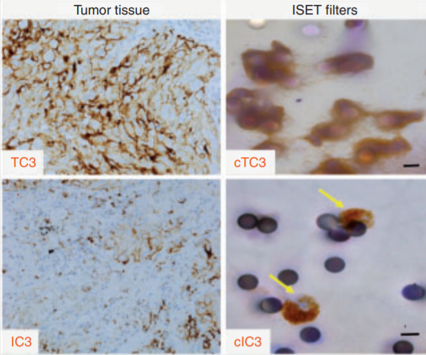
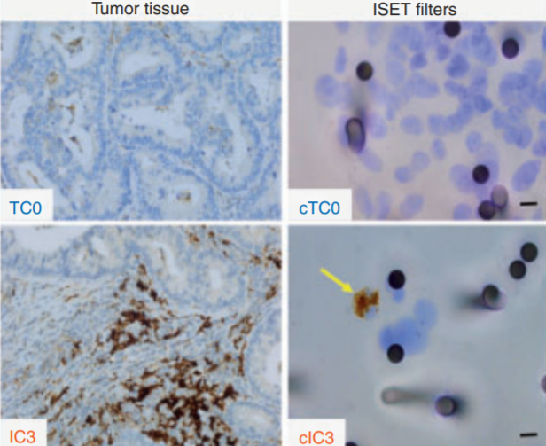
image below shows PD-L1 staining in tumor tissue and corresponding ISET filters from selected NSCLC patients.